

Diagnostic Laparoscopy


Overview
Diagnostic laparoscopy is an operation in which a surgeon makes a few small (typically 5-millimeter) incisions, insufflates the abdomen with carbon dioxide gas, and inserts a thin camera (laparoscope) to look directly at the organs and tissues inside the abdomen and pelvis. It is used when a patient has persistent or unexplained abdominal pain, obstruction, or other symptoms that imaging and endoscopy have failed to diagnose. Its great advantage is that it is both diagnostic and therapeutic in the same setting — once a problem is found (such as an internal hernia, adhesion, or mesenteric defect), it can usually be corrected immediately through the same incisions (Alsulaimy et al., Obesity Surgery, 2017; Facchiano et al., Obesity Surgery, 2016).
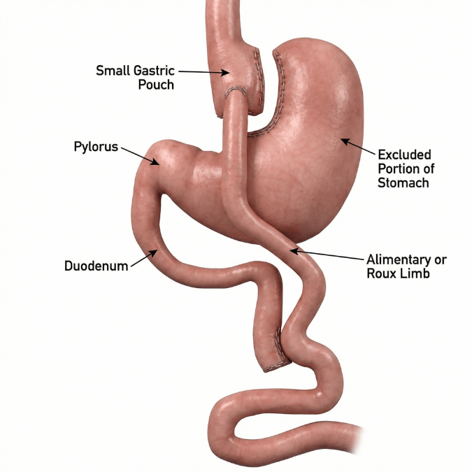
In Dr. de la Cruz-Muñoz's practice, the most important role of diagnostic laparoscopy is in the patient with a prior Roux-en-Y gastric bypass (or other bowel-rerouting operation) who has recurrent crampy, postprandial abdominal pain. Internal hernia is one of the most feared late complications of gastric bypass, and critically, a normal CT scan does not rule it out — abdominal CT has a pooled sensitivity of only about 82%, meaning at least one in six confirmed internal hernias is missed on imaging (Nawas et al., Annals of Surgery, 2022; Fry and Finks, JAMA Surgery, 2023). When clinical suspicion is high, diagnostic laparoscopy becomes the definitive answer — directly inspecting every potential defect and closing any that are found (Oviedo et al., ASMBS, Surgery for Obesity and Related Diseases, 2026).
How the Procedure Works
The procedure works by giving the surgeon a direct, magnified, real-time view of the entire abdominal cavity that no scan can replicate. After the abdomen is inflated with gas to create working space, the laparoscope transmits a high-definition image to monitors in the operating room. The surgeon systematically inspects the abdominal wall, liver, gallbladder, stomach, small and large bowel, mesentery, and pelvic organs. In a patient with altered anatomy from bariatric surgery, the surgeon "runs the bowel" — tracing the intestine from the ileocecal valve backward toward the stomach — to identify kinks, adhesions, intussusception, strictures, or herniation through a mesenteric defect (Alsulaimy et al., Obesity Surgery, 2017; Fry and Finks, JAMA Surgery, 2023). Because the hernia may be intermittent and may have reduced itself by the time of any scan, direct inspection is the only way to reliably confirm or exclude it (Altinoz et al., American Journal of Surgery, 2020)
Benefits
- Highest diagnostic accuracy available — diagnostic laparoscopy is the reference standard for internal hernia and is more accurate than CT, which was falsely negative in 44–50% of patients found to have an internal hernia at surgery (Altinoz et al., American Journal of Surgery, 2020; van Hogezand et al., Surgery for Obesity and Related Diseases, 2026).
- Diagnosis and treatment in one operation — a correctable finding can be repaired immediately, avoiding a second procedure (Facchiano et al., Obesity Surgery, 2016).
- Minimally invasive — small incisions mean less pain, faster recovery, and fewer wound complications than open exploration (Leitman, JAMA Surgery, 2023).
- Relief of chronic pain — when a correctable cause is found and fixed, pain resolves in roughly 70% of patients (Oviedo et al., ASMBS, Surgery for Obesity and Related Diseases, 2026).
- Prevents catastrophe — early exploration can prevent progression of an intermittent internal hernia to bowel strangulation, ischemia, and emergency surgery (Fry and Finks, JAMA Surgery, 2023).
Who Is a Candidate
The ideal candidate is a patient with persistent, recurrent, or unexplained abdominal pain in whom non-invasive testing has been inconclusive — particularly a patient with a prior gastric bypass, SADI, duodenal switch, or one-anastomosis bypass who has recurrent crampy, postprandial pain with or without nausea (Oviedo et al., ASMBS, Surgery for Obesity and Related Diseases, 2026; Alsulaimy et al., Obesity Surgery, 2017). Candidacy is especially clear when imaging shows suspicious signs of internal hernia (mesenteric swirl, venous congestion, mushroom sign), but also applies when imaging is normal yet clinical suspicion remains high (Fry and Finks, JAMA Surgery, 2023; van Hogezand et al., Surgery for Obesity and Related Diseases, 2026). Patients with signs of obstruction or ischemia are candidates for urgent exploration rather than further testing (Fry and Finks, JAMA Surgery, 2023).
How the Procedure Is Performed
The operation is performed under general anesthesia. A common approach in the post-bariatric patient is as follows (Alsulaimy et al., Obesity Surgery, 2017; Kollmann et al., Langenbeck's Archives of Surgery, 2023):
- Safe entry — the abdomen is accessed and insufflated, often at Palmer's point (left upper quadrant) using an optical trocar to avoid injury in patients with prior surgery and adhesions.
- Initial survey — a camera inspects the entry site, abdominal wall, and overall cavity for adhesions or incisional hernias. If dense adhesions are present, additional ports are placed for careful lysis, and conversion to open surgery is considered if safety is in question.
- Running the bowel — the surgeon identifies the cecum and ileocecal valve and traces the small bowel retrograde toward the ligament of Treitz, evaluating the common channel, the entero-enterostomy (jejunojejunostomy), and the alimentary and biliopancreatic limbs for kinking, intussusception, adhesions, or mesenteric defects.
- Inspecting the defects — Petersen's space, the jejunojejunostomy (Brolin) defect, and — in retrocolic reconstructions — the transverse mesocolon defect are each directly examined.
- Repair — any herniated bowel is reduced, viability is assessed (with resection if ischemic), and all open mesenteric defects are closed with permanent (non-absorbable) running suture, taking care not to kink the bowel (Oviedo et al., ASMBS, Surgery for Obesity and Related Diseases, 2026; Kollmann et al., Langenbeck's Archives of Surgery, 2023).
- Additional evaluation — the liver, gallbladder, and pelvic organs are assessed, and upper endoscopy of the pouch and gastrojejunostomy may be performed as indicated (Alsulaimy et al., Obesity Surgery, 2017).
Risks and Considerations
Diagnostic laparoscopy is generally very safe, but like any operation it carries risks, including bleeding, infection, injury to bowel or other organs during entry or adhesiolysis, and the general risks of anesthesia (Alsulaimy et al., Obesity Surgery, 2017). Specific to this setting: a negative exploration (no correctable cause found) is possible and is more likely when imaging is normal, which points toward a functional cause of pain (Oviedo et al., ASMBS, Surgery for Obesity and Related Diseases, 2026; van Hogezand et al., Surgery for Obesity and Related Diseases, 2026). Conversion to open surgery may be necessary if dense adhesions make laparoscopy unsafe (Alsulaimy et al., Obesity Surgery, 2017). When defects are closed, there is a small risk of early bowel obstruction from kinking at the suture site (Oviedo et al., ASMBS, Surgery for Obesity and Related Diseases, 2026).
Recovery Timeline
Recovery after diagnostic laparoscopy is typically rapid because the incisions are small. Many patients go home the same day or after a short observation, and return to normal activity within one to two weeks, depending on what was found and repaired. When a significant repair (such as bowel reduction and defect closure, or resection) is performed, the recovery mirrors that of the therapeutic procedure performed. Patients with acute obstruction or ischemia requiring resection have a longer hospital stay and recovery.
Long-Term Expectations
When a correctable problem such as an internal hernia is found and the defects are closed, the large majority of patients experience lasting relief of chronic pain (Oviedo et al., ASMBS, Surgery for Obesity and Related Diseases, 2026). Patients should understand, however, that defect closure reduces but does not eliminate future risk — mesenteric defects can reopen and internal hernia can recur in up to 40–50% of previously repaired cases, so lifelong awareness of recurrent symptoms remains important (Oviedo et al., ASMBS, Surgery for Obesity and Related Diseases, 2026; Leitman, JAMA Surgery, 2023). For this reason, the mesenteric defects should also be inspected during any future abdominal operation after gastric bypass (Fry and Finks, JAMA Surgery, 2023).
Why Choose Dr. de la Cruz-Muñoz
Dr. Nestor de la Cruz-Muñoz, MD, FACS, DABOM, is a metabolic and bariatric surgeon and board-certified obesity medicine specialist with deep expertise in the altered anatomy of gastric bypass and its long-term complications. Internal hernia is a diagnosis that is routinely missed — nearly one in six are invisible on CT, and the intermittent nature of the pain leads many patients to be told their tests are "normal" (Fry and Finks, JAMA Surgery, 2023; Nawas et al., Annals of Surgery, 2022). Knowing when recurring post-bypass pain warrants diagnostic laparoscopy despite a normal scan, performing a systematic exploration of Petersen's space, the jejunojejunostomy defect, and the transverse mesocolon, and durably closing all defects with permanent suture are precisely the skills that distinguish an experienced bariatric surgeon and that drive symptom relief and lower recurrence (Oviedo et al., ASMBS, Surgery for Obesity and Related Diseases, 2026; Altinoz et al., American Journal of Surgery, 2020). Patients who underwent bariatric or abdominal surgery elsewhere and are now living with unexplained pain are welcome for evaluation.
Total Body Weight Loss (TBWL) Expectations
Not applicable. Diagnostic laparoscopy is an evaluation and repair procedure, not a weight-loss operation, and is not expected to produce weight loss on its own.
Expected Metabolic Improvements
Not applicable. This procedure diagnoses and treats mechanical and structural problems; it does not have an independent metabolic effect.
Weight Regain Prevention Strategy
Not applicable to the diagnostic procedure itself. However, diagnostic laparoscopy is an important tool in the evaluation of patients with post-bariatric symptoms, and findings may inform whether a revisional weight-loss strategy is appropriate (see Revision Options).
GERD / Reflux Considerations
Diagnostic laparoscopy can be a valuable tool in evaluating unexplained foregut symptoms, including reflux, in patients with prior bariatric or anti-reflux surgery, by directly assessing for hiatal hernia, anatomic failure, or other structural causes. Any reflux-specific anatomy identified can often be addressed at the same operation.
Revision Options
Diagnostic laparoscopy frequently serves as the first step that determines whether a revisional or conversion procedure is needed. Findings such as a gastrojejunostomy problem, marginal ulcer, gastro-gastric fistula, stricture, or anatomic failure identified at exploration may lead to a planned revisional operation (Alsulaimy et al., Obesity Surgery, 2017).
Body Composition & Muscle Preservation
Not applicable. This procedure does not affect body composition or muscle mass
Insurance & Qualification Requirements
Diagnostic laparoscopy for abdominal pain, suspected internal hernia, or obstruction is a medically necessary general-surgery procedure and is generally covered by insurance when appropriately indicated. Coverage typically requires documentation of the symptoms, the diagnostic workup performed (including imaging), and the clinical rationale for surgical exploration. Unlike elective bariatric surgery, it does not require a supervised weight-management program.
Preparing for Surgery (Pre-Op Requirements)
Preparation includes a focused history and physical, review of prior operative reports to understand the patient's exact anatomy, and relevant imaging (CT abdomen/pelvis with IV contrast) and/or upper endoscopy as indicated (Nawas et al., Annals of Surgery, 2022; Oviedo et al., ASMBS, Surgery for Obesity and Related Diseases, 2026). Standard preoperative labs, an anesthesia evaluation, and routine fasting instructions apply. Patients should disclose all prior abdominal operations, as this guides safe entry.
Post-Op Nutrition Overview
Diet is advanced according to what is found and repaired. If only inspection or defect closure is performed, patients typically resume a regular (or their established post-bariatric) diet quickly. If bowel is reduced, resected, or an anastomosis is involved, a staged diet advancement is used. Post-bariatric patients should continue their lifelong vitamin and mineral supplementation.
Post-Op Activity & Exercise Guidance
Patients are encouraged to walk the day of surgery to reduce the risk of blood clots. Light activity resumes within days; heavy lifting and strenuous exercise are typically restricted for a short period that depends on the extent of any repair performed. Most patients with a diagnostic-only or defect-closure procedure return to routine activity within one to two weeks.
Special Considerations for International Patients
Quick Procedure Details
Typically covered as a medically necessary diagnostic/therapeutic general-surgery procedure when symptoms and workup support exploration. Prior authorization requirements vary by plan; documentation of failed or non-diagnostic imaging strengthens approval. No bariatric qualification criteria apply.
Featured Procedures




Conditions
FAQ
Because for certain problems — especially internal hernia after gastric bypass — surgery is the only test accurate enough to be trusted. Scans and endoscopy can miss an internal hernia entirely; CT is falsely negative in a large share of confirmed cases because the hernia is often intermittent and may slip back out before imaging (Altinoz et al., American Journal of Surgery, 2020; Nawas et al., Annals of Surgery, 2022). Diagnostic laparoscopy lets the surgeon look directly and, if a problem is found, fix it in the same operation — so it is often both the diagnosis and the cure (Facchiano et al., Obesity Surgery, 2016).
A negative exploration is possible and is more likely when imaging is normal. While it may feel disappointing, it provides valuable information: it makes a structural or surgical cause very unlikely and redirects care toward functional causes of pain — such as dumping syndrome, visceral hypersensitivity, or small intestinal bacterial overgrowth — which are treated medically rather than surgically (Oviedo et al., ASMBS, Surgery for Obesity and Related Diseases, 2026; van Hogezand et al., Surgery for Obesity and Related Diseases, 2026). When a correctable cause is found and fixed, about 70% of patients get lasting pain relief (Oviedo et al., ASMBS, Surgery for Obesity and Related Diseases, 2026).