

Internal Hernia

Videos
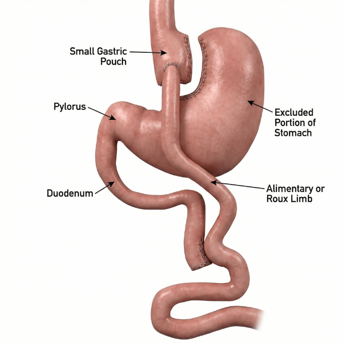
An internal hernia occurs when a loop of small intestine slips through one of the mesenteric openings that are unavoidably created when a surgeon reroutes the bowel during Roux-en-Y gastric bypass (RYGB). Unlike a hernia you can see or feel on the abdominal wall, an internal hernia happens entirely inside the abdomen, where it can twist, obstruct, and cut off the blood supply to the intestine (Fry and Finks, JAMA Surgery, 2023; Wood et al., Radiographics, 2025). It is distinct from — but a leading cause of — post-bariatric small bowel obstruction, and it is one of the single most important reasons any patient with a prior gastric bypass who develops abdominal pain deserves prompt evaluation by a surgeon familiar with altered anatomy (Oviedo et al., ASMBS, Surgery for Obesity and Related Diseases, 2026).
Internal hernia is the complication that makes lifelong surgical follow-up after gastric bypass essential. Roughly half of patients present with acute, severe, sudden-onset pain, while the other half present with chronic, intermittent, milder pain that comes and goes over months or years — a pattern that is easy to dismiss but dangerous to ignore (Oviedo et al., ASMBS, Surgery for Obesity and Related Diseases, 2026; Fry and Finks, JAMA Surgery, 2023).
Internal hernia is caused by small bowel passing through a mesenteric defect — a gap in the fan of tissue that carries blood vessels to the intestine — that forms as a direct consequence of the gastric bypass operation (Murakami et al., Cochrane Database of Systematic Reviews, 2025). There are three classic defect sites, and which ones are present depends on how the bypass was constructed (Fry and Finks, JAMA Surgery, 2023; Desai et al., Abdominal Radiology, 2025):
- Petersen's space (Petersen's defect) — the gap between the mesentery of the Roux (alimentary) limb and the transverse mesocolon. This can form with both antecolic and retrocolic reconstructions.
- Jejunojejunostomy defect ("Brolin's space") — the opening in the small bowel mesentery at the lower connection where the two limbs are joined. This is the most common site of internal hernia.
- Transverse mesocolon defect ("Petersen's hernia" in some references) — present only when the Roux limb is brought up through the colon mesentery in a retrocolic reconstruction. This approach has largely fallen out of favor because of its high internal-hernia rate (Golden et al., Abdominal Radiology, 2024).
Modern antecolic (in front of the colon) reconstruction creates only two defects rather than three, which is one reason technique has shifted over time (Desai et al., Abdominal Radiology, 2025).
- Non-closure of mesenteric defects at the index operation — the single most important modifiable risk factor; leaving defects open raises internal-hernia rates several-fold (Muir et al., Obesity Surgery, 2023; Stenberg et al., JAMA Surgery, 2023).
- Rapid and substantial postoperative weight loss — as mesenteric fat melts away (greatest in the first 1–2 years), defects enlarge and bowel can herniate, which is why most internal hernias present within two years of surgery (Leitman, JAMA Surgery, 2023; Oviedo et al., ASMBS, Surgery for Obesity and Related Diseases, 2026).
- Younger age (Oviedo et al., ASMBS, Surgery for Obesity and Related Diseases, 2026).
- Pregnancy, particularly the second and third trimesters, when the gravid uterus displaces bowel (Oviedo et al., ASMBS, Surgery for Obesity and Related Diseases, 2026).
- Retrocolic Roux limb placement versus antecolic (Golden et al., Abdominal Radiology, 2024; Desai et al., Abdominal Radiology, 2025).
- Laparoscopic (versus open) bypass — fewer post-op adhesions means less of the natural scarring that would otherwise seal defects (Leitman, JAMA Surgery, 2023).
When bowel herniates through a mesenteric defect, it can kink and obstruct, backing up intestinal contents and causing crampy, colicky pain and nausea (Fry and Finks, JAMA Surgery, 2023). If the herniated segment twists on its narrow mesenteric pedicle, it can become a closed-loop obstruction or volvulus, strangling the blood supply. The venous drainage is compressed first — producing the mesenteric congestion and edema seen on imaging — and if unrelieved, arterial inflow is cut off, leading to bowel ischemia and necrosis (Wood et al., Radiographics, 2025; Murakami et al., Cochrane Database of Systematic Reviews, 2025). A critical danger is that the hernia is often intermittent: bowel herniates, causes pain, then slips back out, so the patient feels better and the scan looks normal — until an episode fails to reduce and becomes an emergency (Fry and Finks, JAMA Surgery, 2023).
- Closed-loop obstruction and volvulus, which can progress rapidly (Wood et al., Radiographics, 2025).
- Bowel strangulation, ischemia, necrosis, and perforation — potentially fatal and requiring emergency surgery, sometimes with extensive intestinal resection (Murakami et al., Cochrane Database of Systematic Reviews, 2025).
- Intestinal failure and short bowel syndrome — in catastrophic cases of massive bowel loss, among the most devastating long-term consequences of RYGB, occasionally requiring long-term parenteral nutrition or even visceral transplantation (Vosburg et al., ASMBS Clinical Issues Committee, Surgery for Obesity and Related Diseases, 2026).
- Death — small bowel obstruction following gastric bypass carries a reported mortality of roughly 1–2% (Leitman, JAMA Surgery, 2023).
- Recurrence — even after repair, internal hernia can recur in up to 40–50% of cases from reopened or improperly closed defects (Oviedo et al., ASMBS, Surgery for Obesity and Related Diseases, 2026).
The hallmark symptom is colicky, crampy upper abdominal pain, most commonly after eating (postprandial), frequently accompanied by nausea and vomiting (Oviedo et al., ASMBS, Surgery for Obesity and Related Diseases, 2026). Features associated with higher odds of a true internal hernia include postprandial pain, pain that radiates to the back, localized peritonitis, and an elevated white blood cell count (Oviedo et al., ASMBS, Surgery for Obesity and Related Diseases, 2026). Importantly, the physical exam is often unremarkable and labs may be normal between episodes, which makes the diagnosis notoriously challenging (Fry and Finks, JAMA Surgery, 2023). Red-flag signs of strangulation — severe constant pain, fever, rapid heart rate, and a rigid abdomen — mandate emergency evaluation (Wood et al., Radiographics, 2025).
CT of the abdomen and pelvis with intravenous contrast is the imaging test of choice in a stable patient, with a pooled sensitivity of about 82% and specificity of about 85%, and a positive predictive value of 81% and negative predictive value of 96% (Nawas et al., Annals of Surgery, 2022; Oviedo et al., ASMBS, Surgery for Obesity and Related Diseases, 2026). The most useful CT signs are the mesenteric "swirl" sign (twisting of the mesenteric vessels), superior mesenteric vein "beaking," the "mushroom" sign (herniated mesenteric root), clustered small bowel loops, mesenteric edema, venous congestion, and small bowel positioned behind the superior mesenteric artery (Dilauro et al., Radiology, 2017; Nawas et al., Annals of Surgery, 2022; Yang et al., Abdominal Radiology, 2025).
The crucial caveat — and the point every gastric bypass patient should understand — is that a normal CT scan does not exclude an internal hernia. At least 15% of patients with a surgically confirmed hernia have a normal-appearing scan, both because CT is imperfect and because the intermittent hernia may have reduced itself by the time of imaging (Fry and Finks, JAMA Surgery, 2023; Nawas et al., Annals of Surgery, 2022). For this reason, diagnostic laparoscopy — direct surgical inspection of all mesenteric defects — remains the definitive diagnostic test and may be warranted even when imaging is unremarkable, particularly when pain is severe or obstructive signs are present (Fry and Finks, JAMA Surgery, 2023; Oviedo et al., ASMBS, Surgery for Obesity and Related Diseases, 2026).
In a patient with prior gastric bypass and abdominal pain, the differential includes marginal ulcer, gallstones and biliary colic (common after rapid weight loss), jejunojejunostomy stricture or kinking, intussusception (typically more than two years out), adhesive small bowel obstruction, gastro-gastric fistula, anastomotic leak (early), and functional disorders such as dumping syndrome, small intestinal bacterial overgrowth, and visceral hypersensitivity (Fry and Finks, JAMA Surgery, 2023; Oviedo et al., ASMBS, Surgery for Obesity and Related Diseases, 2026). Excluding marginal ulcer, biliary disease, and anastomotic stricture before attributing symptoms to internal hernia is a standard part of the workup (Oviedo et al., ASMBS, Surgery for Obesity and Related Diseases, 2026).
Internal hernia cannot be prevented by diet or activity changes once the bypass anatomy exists, but patients can protect themselves by understanding the warning signs and acting on them. The most important "lifestyle" measure is behavioral: any gastric bypass patient who develops new, persistent, recurrent, or bilious vomiting or significant abdominal pain should seek evaluation promptly rather than waiting, because delay is what converts a correctable hernia into bowel loss (Oviedo et al., ASMBS, Surgery for Obesity and Related Diseases, 2026). Women who are pregnant or planning pregnancy after bypass should be counseled about the elevated risk during the second and third trimesters and ensure their obstetric team knows their surgical history (Oviedo et al., ASMBS, Surgery for Obesity and Related Diseases, 2026).
Surgical repair is the definitive treatment, and the approach is overwhelmingly minimally invasive (laparoscopic) at experienced centers (Fry and Finks, JAMA Surgery, 2023). The operation involves systematically "running" the bowel — many surgeons start distally at the ileocecal valve and work proximally, where decompressed bowel is easier to trace — reducing any herniated loop, assessing the bowel's viability, and resecting any segment that is ischemic or necrotic (Fry and Finks, JAMA Surgery, 2023). All mesenteric defects are then closed with non-absorbable (permanent) running suture, even when no active herniation is found, because closure both relieves symptoms and prevents recurrence (Oviedo et al., ASMBS, Surgery for Obesity and Related Diseases, 2026).
For the stable patient with chronic, intermittent pain and non-diagnostic imaging, elective diagnostic laparoscopy with defect closure is both diagnostic and therapeutic; when a correctable finding is identified, pain relief rates reach approximately 70%, whereas a negative exploration points toward a functional cause (Oviedo et al., ASMBS, Surgery for Obesity and Related Diseases, 2026). When obstruction or ischemia is suspected, exploration becomes urgent and should not be delayed for additional imaging (Fry and Finks, JAMA Surgery, 2023). Because the risk persists for life, evaluation and closure of these defects is recommended during any future abdominal operation after RYGB — such as a cholecystectomy or hernia repair — even in asymptomatic patients (Fry and Finks, JAMA Surgery, 2023).
The strongest evidence in this field supports prevention: routine closure of all mesenteric defects at the time of the original bypass reduces internal-hernia risk by roughly 70% (Muir et al., Obesity Surgery, 2023; Oviedo et al., ASMBS, Surgery for Obesity and Related Diseases, 2026). In the landmark Swedish randomized trial of 2,507 patients, defect closure nearly halved reoperations for small bowel obstruction over 10 years (7.8% with closure versus 14.9% without), which is why closure is now considered standard practice (Stenberg et al., JAMA Surgery, 2023; Leitman, JAMA Surgery, 2023).
Any patient with a prior gastric bypass who develops recurring crampy abdominal pain, especially after meals, with or without nausea — even if episodes come and go and even if a previous scan was normal — should be evaluated by a bariatric surgeon familiar with their anatomy (Fry and Finks, JAMA Surgery, 2023; Oviedo et al., ASMBS, Surgery for Obesity and Related Diseases, 2026). Chronic, intermittent symptoms should never be brushed off as "normal after bypass."
Seek emergency care immediately for severe, constant abdominal pain, repeated vomiting (particularly bilious vomiting), inability to pass gas or stool, a distended or rigid abdomen, fever, rapid heart rate, or lightheadedness — these may signal strangulation, closed-loop obstruction, or ischemia requiring emergency surgery (Wood et al., Radiographics, 2025; Murakami et al., Cochrane Database of Systematic Reviews, 2025). In bypass patients, the window to save the bowel can be short, so timely surgical exploration is prioritized over prolonged observation (Fry and Finks, JAMA Surgery, 2023).
The long-term outlook is favorable with modern technique and vigilant follow-up. Routine closure of all defects at the index operation has measurably reduced internal-hernia and obstruction rates over time (Stenberg et al., JAMA Surgery, 2023; Muir et al., Obesity Surgery, 2023). Patients should understand, however, that closure reduces but does not eliminate the risk — defects can reopen, and hernia can recur in 40–50% of previously repaired cases — so lifelong awareness of symptoms and lifelong bariatric follow-up remain essential (Leitman, JAMA Surgery, 2023; Oviedo et al., ASMBS, Surgery for Obesity and Related Diseases, 2026). With prompt attention to recurring pain, the overwhelming majority of patients return to their expected post-bypass trajectory.
With timely diagnosis and laparoscopic repair, most patients recover well and the great majority of internal hernias are managed minimally invasively at experienced centers (Leitman, JAMA Surgery, 2023). Prognosis hinges almost entirely on how quickly the hernia is recognized and treated: when caught before strangulation, outcomes are excellent, but delayed diagnosis with bowel ischemia or perforation carries substantial morbidity and a mortality of roughly 1–2% for post-bypass obstruction (Leitman, JAMA Surgery, 2023; Murakami et al., Cochrane Database of Systematic Reviews, 2025). When a correctable defect is found and closed, chronic pain resolves in the large majority of patients (Oviedo et al., ASMBS, Surgery for Obesity and Related Diseases, 2026).
Dr. Nestor de la Cruz-Muñoz, MD, FACS, DABOM, is a metabolic and bariatric surgeon and board-certified obesity medicine specialist with deep expertise in the altered anatomy of gastric bypass and its long-term complications. Internal hernia is a diagnosis that is routinely missed — nearly one in six are invisible on CT, and the intermittent nature of the pain leads many patients to be told their tests are "normal" (Fry and Finks, JAMA Surgery, 2023; Nawas et al., Annals of Surgery, 2022). Recognizing when recurring post-bypass pain warrants diagnostic laparoscopy despite a normal scan, performing a systematic exploration of Petersen's space, the jejunojejunostomy defect, and the transverse mesocolon, and durably closing all defects with permanent suture are precisely the skills that distinguish an experienced bariatric surgeon and that are most strongly associated with symptom relief and lower recurrence (Oviedo et al., ASMBS, Surgery for Obesity and Related Diseases, 2026; Stenberg et al., JAMA Surgery, 2023). Patients who underwent bypass elsewhere and are now living with unexplained abdominal pain are welcome for evaluation.
Featured Procedures



FAQ
Yes. Internal hernia is a risk after essentially any bariatric operation that reroutes the small bowel and creates a mesenteric defect — not just the classic Roux-en-Y gastric bypass. It has been documented after one-anastomosis (mini) gastric bypass (OAGB), single-anastomosis duodeno-ileal bypass with sleeve (SADI-S), and biliopancreatic diversion with duodenal switch (BPD/DS) (Khogeer et al., Obesity Surgery, 2021; Davey et al., Obesity Surgery, 2026). What differs is how often it happens, and that difference is driven by how many mesenteric defects each operation creates.
Operations with a single anastomosis and fewer (or essentially unclosed, wide-open) mesenteric defects tend to have lower internal hernia rates. In a network meta-analysis of randomized trials, reported internal hernia rates were about 2.4% for Roux-en-Y gastric bypass, 3.1% for OAGB, 1.8% for duodenal switch, and 0% for SADI-S, with no statistically significant difference between them (Davey et al., Obesity Surgery, 2026). Procedures with an extra bowel connection carry a higher risk: the additional ileo-ileal anastomosis in traditional BPD/DS creates more mesenteric defects, and internal hernia consistently occurs more often after BPD/DS than after the single-anastomosis SADI-S (roughly 8% vs 1.5% in pooled series) (Galvao Goncalves et al., Surgical Endoscopy, 2026; El Ghazal et al., Surgical Endoscopy, 2025). OAGB was once thought to almost never cause internal hernia, but larger series have shown it is more common — and more often missed — than previously believed, so a low threshold for evaluation applies regardless of which bypass a patient had (Petrucciani et al., Obesity Surgery, 2021; Facchiano et al., Obesity Surgery, 2020).
The practical takeaway: any patient with a rerouted gut — OAGB, SADI, DS, or classic gastric bypass — who develops recurrent crampy, postprandial abdominal pain deserves prompt evaluation by a surgeon familiar with their specific anatomy, because the pain can be intermittent and the CT scan can look normal (Khogeer et al., Obesity Surgery, 2021).
No — a normal CT scan does not rule out an internal hernia. This is one of the most important and least understood facts for anyone living with a bypass. Pooled data show abdominal CT has a sensitivity of only about 82%, meaning roughly one in six patients with a surgically confirmed internal hernia will have a scan that looks normal (Nawas et al., Annals of Surgery, 2022; Fry and Finks, JAMA Surgery, 2023). There are two reasons: CT is an imperfect test, and internal hernias are frequently intermittent — the bowel herniates and causes pain, then slips back out before the scan is taken, so the imaging is captured during a "quiet" window.
Because of this, the decision to operate is based on the whole clinical picture — recurrent postprandial crampy pain, nausea, and the pattern of symptoms — not on imaging alone. When suspicion is high despite a normal scan, diagnostic laparoscopy (directly inspecting all the mesenteric defects) remains the definitive test and is both diagnostic and therapeutic (Fry and Finks, JAMA Surgery, 2023; Oviedo et al., ASMBS, Surgery for Obesity and Related Diseases, 2026). If a correctable finding is identified and the defect closed, pain resolves in roughly 70% of patients (Oviedo et al., ASMBS, Surgery for Obesity and Related Diseases, 2026). The dangerous mistake is to be reassured by a single normal scan and wait — delay is what allows an intermittent, correctable hernia to progress to bowel strangulation and an emergency.