

Small Bowel Obstruction (SBO)

Videos
Small bowel obstruction occurs when the normal flow of intestinal contents is mechanically blocked, causing fluid and gas to accumulate proximal to the blockage. This distension raises pressure within the bowel wall, reduces mucosal blood flow, and — if unrelieved — can progress to ischemia, necrosis, and perforation (Rami Reddy and Cappell, Current Gastroenterology Reports, 2017). It is one of the most common surgical emergencies, accounting for roughly 350,000 admissions per year in the United States and more than 60% of emergency laparotomies in some health systems (Rami Reddy and Cappell, Current Gastroenterology Reports, 2017; Green et al., European Journal of Trauma and Emergency Surgery, 2023).
This page covers SBO in two distinct contexts that Dr. de la Cruz-Muñoz manages: the general surgical patient, where adhesions and hernias dominate, and the bariatric/metabolic surgery patient, where internal hernias, anastomotic strictures, and other procedure-specific causes change both the diagnosis and the urgency of treatment (Wernick et al., European Journal of Trauma and Emergency Surgery, 2016; De Simone et al., OBA Guidelines, 2022).
General surgical causes. In the overall population, adhesions from prior abdominal or pelvic surgery are by far the most common cause (~65%), followed by hernias (~10%), neoplasms (~5%), Crohn's disease (~5%), and other causes (~15%) (Rami Reddy and Cappell, Current Gastroenterology Reports, 2017). Adhesions and hernias together account for roughly 90% of mechanical SBO (Alashaby et al., Scientific Reports, 2026). Laparoscopic surgery is associated with fewer adhesions than open surgery because it causes less peritoneal trauma and fibrin deposition (Abera et al., Scientific Reports, 2025).
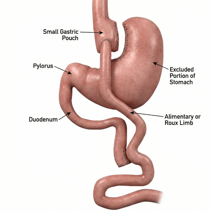
Bariatric surgery causes. After Roux-en-Y gastric bypass (RYGB), the causes are different and frequently require operative correction. The leading cause is internal hernia (>50% of late obstructions), which can occur at three classic sites: Petersen's space, the jejunojejunostomy mesenteric defect, and the transverse mesocolon defect (in retrocolic reconstructions) (Chand and Prathanvanich, Journal of Intensive Care Medicine, 2016; McLean et al., Obesity Surgery, 2024). Other bariatric-specific causes include jejunojejunostomy stenosis or kinking, adhesions, incisional/trocar-site hernia, intussusception, volvulus, bezoar, and intraluminal blood clot (De Simone et al., OBA Guidelines, 2022; Gabrielli et al., Langenbeck's Archives of Surgery, 2021). Early postoperative obstruction is more often due to anastomotic edema or hematoma, whereas late obstruction (weeks to months out) is more often internal hernia or stricture (Wernick et al., European Journal of Trauma and Emergency Surgery, 2016; Kumbhari et al., AGA Clinical Practice Update, 2021).
- Prior abdominal or pelvic surgery — the dominant risk factor for adhesive SBO (Baiu and Hawn, JAMA, 2018).
- Abdominal wall or inguinal hernia (Baiu and Hawn, JAMA, 2018).
- Roux-en-Y gastric bypass — carries an ~11-fold higher odds of SBO compared with sleeve gastrectomy, making it the largest procedure-related predictor among elective bariatric cases (McLean et al., Obesity Surgery, 2024).
- Other Bypass operations — such as the One Anastomosis Gastric Bypass, Duodenal Switch, SADi-S, and MagDI+Sleeve
- Retrocolic Roux limb placement and non-closure of mesenteric defects — raise internal-hernia risk versus antecolic placement with defect closure (Gabrielli et al., Langenbeck's Archives of Surgery, 2021; McLean et al., Obesity Surgery, 2024).
- Lower BMI, older age, female sex, and significant weight loss after bypass — a thinner mesentery and larger mesenteric defects predispose to internal herniation (McLean et al., Obesity Surgery, 2024).
- Prior history of SBO, intra-abdominal malignancy, Crohn's disease (Rami Reddy and Cappell, Current Gastroenterology Reports, 2017).
Proximal to the blockage, the bowel distends with swallowed air and accumulating intestinal fluid. Rising intraluminal pressure stretches the bowel wall, impairing mucosal perfusion and allowing bacterial overgrowth and decreased wall integrity (Rami Reddy and Cappell, Current Gastroenterology Reports, 2017). Vomiting and third-spacing of fluid into the bowel lumen cause dehydration and electrolyte disturbances. In a closed-loop obstruction or when a segment is strangulated, blood supply is cut off and the bowel can rapidly become ischemic and perforate — the sequence that drives SBO mortality (Alashaby et al., Scientific Reports, 2026). A critical pitfall in bypass patients is that the excluded gastric remnant and biliopancreatic limb cannot be decompressed by a nasogastric tube, so the bowel upstream of the obstruction remains under pressure (Chand and Prathanvanich, Journal of Intensive Care Medicine, 2016).
- Bowel ischemia, necrosis, and perforation from strangulation or closed-loop obstruction (Rami Reddy and Cappell, Current Gastroenterology Reports, 2017).
- Sepsis and peritonitis, with overall SBO mortality ~10%, rising to ~30% with necrosis or perforation (Rami Reddy and Cappell, Current Gastroenterology Reports, 2017).
- Dehydration, electrolyte derangement, and acute kidney injury.
- In bariatric patients specifically — higher rates of anastomotic leak, reoperation, surgical-site infection, readmission, and ICU stay when SBO occurs (McLean et al., Obesity Surgery, 2024).
- Recurrence — particularly after nonoperatively managed adhesive SBO, where recurrence reaches ~24% versus ~9% after surgical management (Chierici et al., Annals of Surgery, 2026).
The classic tetrad is cramping abdominal pain, nausea and vomiting, abdominal distension, and constipation progressing to obstipation (Rami Reddy and Cappell, Current Gastroenterology Reports, 2017). Physical findings may include abdominal distension, abnormal or high-pitched bowel sounds, and signs of dehydration. Warning signs of strangulation include fever, tachycardia, hypotension, diffuse pain, peritonitis, and laboratory findings of marked leukocytosis, neutrophilia, or lactic acidosis (Long et al., Journal of Emergency Medicine, 2019).
A key caveat in post-bypass patients: because the gastric pouch is small, vomiting is often absent, and bilious vomiting, when present, points to obstruction at the jejunojejunostomy. Any bypass patient with persistent abdominal pain — even with unremarkable imaging — must be taken seriously for internal hernia (De Simone et al., OBA Guidelines, 2022).
Diagnosis combines history (prior surgery, hernia, cancer), physical exam, and imaging. CT of the abdomen and pelvis with contrast is the imaging modality of choice, with roughly 90% sensitivity and specificity, identifying the level, cause, and severity of obstruction and signs of ischemia (Green et al., European Journal of Trauma and Emergency Surgery, 2023; Rami Reddy and Cappell, Current Gastroenterology Reports, 2017). Plain radiographs may show dilated loops and air-fluid levels but cannot exclude SBO (Long et al., Journal of Emergency Medicine, 2019). Water-soluble (Gastrografin) contrast studies both help predict which obstructions will resolve nonoperatively and may have a therapeutic effect (Alverdy et al., JAMA Surgery, 2026).
In bypass patients, the radiologist must actively look for internal hernia; the single best CT sign is the "mesenteric swirl," with added accuracy when combined with a "mushroom"-shaped mesenteric root — findings that can be present even before complete obstruction and should prompt early surgical exploration (Riaz et al., Abdominal Radiology, 2016).
Key alternatives include postoperative ileus, opioid-induced (narcotic) bowel dysfunction, colonic pseudo-obstruction (Ogilvie syndrome), large bowel obstruction, and mesenteric ischemia (Rami Reddy and Cappell, Current Gastroenterology Reports, 2017). In bariatric patients, marginal ulcer, anastomotic leak, gastro-gastric fistula, and gastric sleeve stenosis/kinking should also be considered (De Simone et al., OBA Guidelines, 2022; Chand and Prathanvanich, Journal of Intensive Care Medicine, 2016).
SBO is largely not prevented by lifestyle measures, but general strategies include maintaining a healthy weight, promptly repairing symptomatic hernias before incarceration, and — for patients with a known adhesion or prior SBO history — seeking care early when obstructive symptoms begin rather than waiting (Baiu and Hawn, JAMA, 2018). Bariatric patients should be counseled to present immediately for new, persistent, or bilious vomiting or severe abdominal pain, because delay in internal-hernia diagnosis risks bowel loss (De Simone et al., OBA Guidelines, 2022).
General surgery approach. For uncomplicated adhesive SBO without peritonitis, strangulation, or ischemia, initial management is nonoperative: bowel rest (nil per os), intravenous fluid resuscitation, electrolyte correction, and nasogastric decompression, with serial exams over a 48–72-hour trial (Alverdy et al., JAMA Surgery, 2026; Green et al., European Journal of Trauma and Emergency Surgery, 2023). Nonoperative management resolves roughly 70–90% of adhesive episodes (Abera et al., Scientific Reports, 2025). Surgery — lysis of adhesions, preferentially laparoscopic, with resection of any nonviable bowel — is required for strangulation, closed-loop obstruction, perforation, or failure of nonoperative therapy (Baiu and Hawn, JAMA, 2018; Rami Reddy and Cappell, Current Gastroenterology Reports, 2017). Timing matters: delayed surgery is associated with higher early mortality (3.6% within 0–1 days vs 7.1% after ≥9 days) and more bowel resection, while surgical management — especially laparoscopic — substantially reduces long-term recurrence (Chierici et al., Annals of Surgery, 2026).
Bariatric surgery considerations. The threshold for operation is much lower after RYGB. Nasogastric decompression is frequently ineffective because it cannot reach the biliopancreatic limb or excluded remnant, and prolonged nonoperative management can be dangerous (Chand and Prathanvanich, Journal of Intensive Care Medicine, 2016). Guidelines advise that in a stable bypass patient with persistent pain and inconclusive imaging, exploratory laparoscopy within 12–24 hours is mandatory to exclude internal hernia (De Simone et al., OBA Guidelines, 2022). Exploration should run the bowel systematically to inspect Petersen's space, the jejunojejunostomy mesenteric defect, and (in retrocolic cases) the transverse mesocolon; viable hernias are reduced and defects closed with non-absorbable suture, while ischemic bowel is resected (De Simone et al., OBA Guidelines, 2022). That said, select cases of early post-bypass obstruction — often from edema or temporary pathology at the jejunojejunostomy — can resolve nonoperatively in carefully monitored, stable patients without alarm signs (up to ~80% in one recent series) (Lazar et al., Obesity Surgery, 2025). Endoscopic therapy (pneumatic dilation of strictures, or stenting for select acute anastomotic obstructions) is an additional option in appropriate bariatric cases (Kumbhari et al., AGA Clinical Practice Update, 2021; De Simone et al., OBA Guidelines, 2022).
Anyone with persistent cramping abdominal pain, repeated vomiting, inability to pass gas or stool, or a progressively distending, bloated abdomen should seek urgent medical evaluation — especially with a history of prior abdominal surgery or a hernia (Baiu and Hawn, JAMA, 2018). Bariatric surgery patients with any persistent or bilious vomiting or significant abdominal pain should be evaluated promptly, as symptoms may be the only early sign of a correctable internal hernia (De Simone et al., OBA Guidelines, 2022).
Anyone with persistent cramping abdominal pain, repeated vomiting, inability to pass gas or stool, or a progressively distending, bloated abdomen should seek urgent medical evaluation — especially with a history of prior abdominal surgery or a hernia (Baiu and Hawn, JAMA, 2018). Bariatric surgery patients with any persistent or bilious vomiting or significant abdominal pain should be evaluated promptly, as symptoms may be the only early sign of a correctable internal hernia (De Simone et al., OBA Guidelines, 2022).
Surgical management reduces long-term recurrence compared with repeated nonoperative treatment, and laparoscopic approaches lower recurrence further (Chierici et al., Annals of Surgery, 2026). For bariatric patients, modern technique — antecolic Roux reconstruction with routine closure of all mesenteric defects — has measurably reduced the rate of internal-hernia obstruction over time, and patients who undergo timely repair generally return to their expected postoperative trajectory (Gabrielli et al., Langenbeck's Archives of Surgery, 2021; McLean et al., Obesity Surgery, 2024).
Most adhesive SBO episodes resolve with nonoperative management, and outcomes have improved with CT-guided decision-making and minimally invasive surgery (Green et al., European Journal of Trauma and Emergency Surgery, 2023). Overall mortality is about 10%, but climbs to roughly 30% when bowel necrosis or perforation has occurred — underscoring that prognosis hinges on early recognition and timely intervention (Rami Reddy and Cappell, Current Gastroenterology Reports, 2017). In bariatric patients, SBO is uncommon (~0.3–6% depending on procedure and era) but carries meaningful morbidity when it occurs, with the great majority of internal-hernia cases now manageable laparoscopically at experienced centers (McLean et al., Obesity Surgery, 2024; Gabrielli et al., Langenbeck's Archives of Surgery, 2021).
Dr. Nestor de la Cruz-Muñoz, MD, FACS, DABOM, is a metabolic and bariatric surgeon and board-certified obesity medicine specialist whose practice spans both general surgical emergencies and the specialized care of post-bariatric complications. This dual expertise is critical for small bowel obstruction: distinguishing a routine adhesive obstruction that may resolve with observation from a post-bypass internal hernia that demands urgent laparoscopic exploration requires surgeons familiar with altered anatomy and the specific imaging signs it produces (De Simone et al., OBA Guidelines, 2022; Riaz et al., Abdominal Radiology, 2016). His emphasis on minimally invasive technique, systematic exploration, and routine mesenteric-defect closure reflects the strategies most strongly associated with lower recurrence and better outcomes (Gabrielli et al., Langenbeck's Archives of Surgery, 2021; Chierici et al., Annals of Surgery, 2026).
Featured Procedures






FAQ
Yes. One of the most important things for gastric bypass patients to understand is that an internal hernia — the leading cause of late obstruction after Roux-en-Y gastric bypass — often does not present as a dramatic, all-at-once blockage. It frequently causes chronic, vague, crampy, postprandial abdominal pain and nausea that comes and goes over weeks, months, or even years, because the bowel repeatedly herniates through a mesenteric defect and then slips back out (Fry and Finks, JAMA Surgery, 2023; Zevallos and Pouwels, Surgical Endoscopy, 2026). This intermittent nature is exactly what makes it dangerous: the hernia may spontaneously reduce by the time imaging is obtained, so a CT scan performed between episodes can look completely normal — yet at least 15% of patients with a true internal hernia have a normal CT even during symptoms (Fry and Finks, JAMA Surgery, 2023). Other causes of chronic, waxing-and-waning obstruction after bypass include adhesions, jejunojejunostomy stricture or kinking with chronic dilation of the biliopancreatic or Roux limb, and intussusception (which typically appears more than two years out) (Wernick et al., European Journal of Trauma and Emergency Surgery, 2016; Fry and Finks, JAMA Surgery, 2023). Because biliopancreatic limb obstruction in particular produces nonspecific, intermittent pain often unrelated to eating, any bypass patient with recurrent unexplained abdominal pain deserves evaluation by a bariatric surgeon — and a normal scan should never be the end of the workup (Dar et al., Obesity Surgery, 2020; De Simone et al., OBA Guidelines, 2022).
Absolutely, and this is one of the most dangerous misconceptions after Roux-en-Y gastric bypass. A normal CT scan does not rule out an internal hernia. CT is only about 82% sensitive, meaning roughly one in six patients with a true hernia will have a normal-appearing scan — and because the hernia is often intermittent, it may have reduced itself by the time the scan is performed (Fry and Finks, JAMA Surgery, 2023; Wood et al., Radiographics, 2025). The most useful warning signs the radiologist looks for are the "mesenteric swirl" sign, venous congestion, and mesenteric edema, but their absence is not reassuring in a patient with persistent symptoms (Fry and Finks, JAMA Surgery, 2023). For this reason, international guidelines recommend that a stable bypass patient with persistent abdominal pain and inconclusive labs and imaging undergo diagnostic laparoscopy — even with a normal CT — to directly inspect Petersen's space, the jejunojejunostomy mesenteric defect, and the transverse mesocolon (De Simone et al., OBA Guidelines, 2022; Chand and Prathanvanich, Journal of Intensive Care Medicine, 2016). Elective laparoscopic closure of a symptomatic mesenteric defect resolves the chronic pain in nearly all such patients, and this is why experienced bariatric surgeons maintain a low threshold to explore rather than dismiss recurring symptoms (Vosburg et al., ASMBS Clinical Issues Committee Review, 2026). If you have had a gastric bypass and are living with recurrent pain and bloating despite "normal" tests, seek evaluation from a bariatric surgeon familiar with your altered anatomy.