
Erosive (Reflux) Esophagitis

Videos
Erosive esophagitis (EE) is the phenotype of gastroesophageal reflux disease in which reflux of acid and other gastric contents produces visible breaks — erosions — in the mucosa of the distal esophagus that can be seen directly at upper endoscopy (Maret-Ouda et al., JAMA, 2020). It is one of three recognized GERD phenotypes, alongside non-erosive reflux disease and Barrett's esophagus, and it accounts for roughly 30% of patients with typical reflux symptoms (Spechler et al., Clinical Gastroenterology and Hepatology, 2024). Esophagitis is the most common complication of GERD, developing in about 18–25% of patients with reflux symptoms (Maret-Ouda et al., JAMA, 2020).
The defining feature of erosive esophagitis — and what separates it clinically from non-erosive reflux disease — is that the mucosal damage is visible and objective. The findings of erosive esophagitis are specific for the diagnosis of GERD, which means that in most cases EE confirms reflux disease without the need for further acid testing (Katz et al., ACG Clinical Guideline, 2022). Severity is scored by the Los Angeles (LA) classification, the most widely used and validated system, graded from A (minimal) to D (very severe) by the extent of the mucosal breaks (Maret-Ouda et al., JAMA, 2020). This grading is not academic: it directly drives treatment duration, the need for follow-up endoscopy, and the likelihood of recurrence — higher grades heal more slowly, relapse faster, and demand indefinite therapy (Katz et al., ACG Clinical Guideline, 2022). Because erosive esophagitis is reliably healed by acid suppression, a diagnosis of non-erosive reflux disease should only be made when endoscopy is performed off PPIs, since ongoing therapy can mask underlying EE (see our Non-Erosive Reflux Disease page) (Katz et al., ACG Clinical Guideline, 2022).
Erosive esophagitis is caused by the reflux of injurious gastric contents — acid, pepsin, and bile — into the esophagus in amounts or for durations the esophagus cannot clear or neutralize (Mittal and Vaezi, New England Journal of Medicine, 2020). Several mechanisms converge to produce visible erosions:
- Prolonged esophageal acid exposure, often driven by a mechanically incompetent esophagogastric junction — a hypotensive lower esophageal sphincter, a hiatal hernia, supine (nighttime) reflux, and poor acid clearance — which is especially characteristic of high-grade (LA C and D) disease (Katzka et al., Clinical Gastroenterology and Hepatology, 2020).
- The combined injury of acid and pepsin, which is more damaging to the esophageal epithelium than acid alone; pepsin activity falls sharply once the pH rises above 4, which is why acid suppression is so effective (Mittal and Vaezi, New England Journal of Medicine, 2020).
- An inflammatory, immune-mediated response. Contrary to the older "acid burn" model, the erosions appear to result from a cytokine- and T-cell-mediated inflammatory reaction triggered by the refluxate, rather than simple chemical cautery of the surface cells (Argüero and Sifrim, Nature Reviews Gastroenterology & Hepatology, 2024).
- Impaired mucosal defenses, including reduced salivary bicarbonate (as seen in xerostomia and Sjögren's syndrome) and dilated intercellular spaces that increase epithelial permeability (Mittal and Vaezi, New England Journal of Medicine, 2020).
A large meta-analysis of more than 759,000 participants identified the factors that most increase the risk of erosive esophagitis (Witarto et al., Scientific Reports, 2023):
- Hiatal hernia — the strongest single risk factor (odds ratio ~4.07) (see our Hiatal & Paraesophageal Hernia page).
- Obesity, both general (OR ~1.78) and central/abdominal, and the related metabolic conditions — diabetes, hypertension, dyslipidemia, hypertriglyceridemia, and non-alcoholic fatty liver disease.
- Age 60 or older (OR ~2.03).
- White/Caucasian ethnicity (OR ~1.67) and male sex, particularly for high-grade disease and asymptomatic EE (Katzka et al., Clinical Gastroenterology and Hepatology, 2020).
- Long-standing GERD (symptoms for 5 years or more).
Notably, Helicobacter pylori infection and atrophic gastritis are protective against erosive esophagitis, because they reduce gastric acid output (Witarto et al., Scientific Reports, 2023).
In erosive esophagitis, repeated exposure of the distal esophageal lining to acid and pepsin triggers an inflammatory cascade that breaks down the protective squamous epithelium, producing the visible erosions that define the condition (Argüero and Sifrim, Nature Reviews Gastroenterology & Hepatology, 2024). The loss of surface cells stimulates basal cell hyperplasia, and inflammatory mediators activate sensory nerve endings, generating the sensation of heartburn (Mittal and Vaezi, New England Journal of Medicine, 2020). Importantly, symptom severity correlates poorly with the degree of visible damage — between 11% and 47% of patients with erosive esophagitis report neither heartburn nor regurgitation, a phenomenon more common in older men and those with higher body mass index (Katzka et al., Clinical Gastroenterology and Hepatology, 2020). High-grade disease reflects largely irreversible mechanical defects of the esophagogastric junction, which is why it tends to require open-ended therapy unless addressed with profound weight loss or antireflux surgery (Katzka et al., Clinical Gastroenterology and Hepatology, 2020). Left unhealed, chronic erosion and the repair that follows can lead to fibrosis and stricture, and to the metaplastic change of Barrett's esophagus.
The complications of erosive esophagitis arise from chronic, unhealed mucosal injury (Richter and Rubenstein, Gastroenterology, 2018):
- Esophageal stricture — fibrotic narrowing of the esophagus causing difficulty swallowing, historically affecting roughly 0.1% of reflux patients; stricture-related hospitalizations and the need for repeat dilation have fallen with widespread PPI use (Richter and Rubenstein, Gastroenterology, 2018; Moayyedi and Talley, Lancet, 2006).
- Gastrointestinal bleeding from the eroded mucosa, which can cause anemia, especially in severe (LA grade D) disease (Richter and Rubenstein, Gastroenterology, 2018).
- Esophageal ulcer (prevalence ~0.05%) (Moayyedi and Talley, Lancet, 2006).
- Barrett's esophagus — a metaplastic change from squamous to columnar mucosa associated with chronic, severe reflux; it is the precursor lesion to esophageal adenocarcinoma, which develops in roughly 0.5–1% of Barrett's patients per year (see our Barrett's Esophagus page) (Moayyedi and Talley, Lancet, 2006).
- Esophageal adenocarcinoma — the most feared, though uncommon, long-term consequence of chronic erosive reflux (Maret-Ouda et al., JAMA, 2020).
Reassuringly, most patients with erosive esophagitis follow a benign course: in a natural-history study, 73–82% of patients had healed esophagitis at follow-up endoscopy, and only a minority progressed to Barrett's esophagus regardless of grade, gender, or continued PPI use (Bi et al., Digestive Diseases and Sciences, 2020).
The symptoms of erosive esophagitis are the typical symptoms of reflux disease, though a substantial minority of patients are asymptomatic (Katzka et al., Clinical Gastroenterology and Hepatology, 2020):
- Heartburn — burning discomfort behind the breastbone, the hallmark symptom.
- Regurgitation of acid or food into the throat or mouth.
- Chest pain (non-cardiac).
- Dysphagia (difficulty swallowing) — which may signal a stricture and always warrants evaluation (Richter and Rubenstein, Gastroenterology, 2018).
- Odynophagia (painful swallowing).
- Signs of bleeding — anemia, or overt gastrointestinal bleeding, in severe disease (Richter and Rubenstein, Gastroenterology, 2018).
Alarm features — dysphagia, unintentional weight loss, gastrointestinal bleeding or anemia, or vomiting — require prompt endoscopy (Katz et al., ACG Clinical Guideline, 2022). Because 11–47% of patients have no typical symptoms at all, erosive esophagitis is sometimes discovered incidentally at endoscopy performed for other reasons (Katzka et al., Clinical Gastroenterology and Hepatology, 2020).
Erosive esophagitis is a diagnosis made at endoscopy, and in most cases the finding alone establishes GERD without additional acid testing (Katz et al., ACG Clinical Guideline, 2022):
- Upper endoscopy (EGD) is the diagnostic test — it directly visualizes the mucosal breaks and allows grading (Katz et al., ACG Clinical Guideline, 2022).
- Los Angeles (LA) classification grades severity: grade A (one or more breaks ≤5 mm), grade B (one or more breaks >5 mm), grade C (breaks bridging the tops of ≥2 mucosal folds, involving <75% of the circumference), and grade D (breaks involving ≥75% of the circumference) (Maret-Ouda et al., JAMA, 2020).
- Diagnostic significance by grade: LA grade A is not sufficient on its own to diagnose GERD because it cannot be reliably distinguished from normal; LA grade B is diagnostic in the presence of typical symptoms and PPI response; LA grades C and D are virtually always diagnostic of GERD (Katz et al., ACG Clinical Guideline, 2022).
- Follow-up endoscopy is recommended after PPI treatment in LA grade C and D disease, both to confirm healing and to evaluate for underlying Barrett's esophagus, which can be hidden by severe inflammation (Katz et al., ACG Clinical Guideline, 2022).
- Ambulatory reflux (pH) testing is generally not needed to confirm GERD once erosive esophagitis is established, but is reserved for the overlap with non-erosive and functional disease (see our Non-Erosive Reflux Disease and PPI-Refractory GERD pages) (Katz et al., ACG Clinical Guideline, 2022).
Several conditions can mimic erosive esophagitis or cause esophageal mucosal breaks and must be distinguished (Fass, New England Journal of Medicine, 2022):
- Non-erosive reflux disease (NERD) — reflux symptoms with a normal endoscopy, the most common GERD phenotype (see our Non-Erosive Reflux Disease page).
- Barrett's esophagus — metaplastic columnar mucosa, which may coexist with or be masked by severe esophagitis (see our Barrett's Esophagus page).
- Eosinophilic esophagitis — an immune/allergic esophageal disorder diagnosed on biopsy, which can produce similar symptoms and a mucosa that looks inflamed.
- Pill-induced (medication) esophagitis — from agents such as bisphosphonates, tetracyclines, NSAIDs, or potassium chloride lodging in the esophagus.
- Infectious esophagitis — Candida, herpes simplex virus, or cytomegalovirus, particularly in immunocompromised patients.
- Achalasia and other motility disorders — which can cause retention esophagitis and mimic reflux (see our Achalasia page).
Lifestyle measures are a foundational part of management, used alongside — not instead of — acid-suppressing therapy in erosive disease (Fass, New England Journal of Medicine, 2022):
- Weight loss in overweight and obese patients — the measure with the strongest supporting evidence, given the central role of obesity and hiatal hernia in EE (Witarto et al., Scientific Reports, 2023).
- Avoiding meals within 3 hours of bedtime and elevating the head of the bed, which is especially relevant because high-grade esophagitis is driven by supine, nighttime reflux (Thrumurthy et al., JAMA, 2026; Katzka et al., Clinical Gastroenterology and Hepatology, 2020).
- Smoking cessation and reducing alcohol (Fass, New England Journal of Medicine, 2022).
- Avoiding individual trigger foods.
These measures reduce reflux burden but do not correct a mechanical problem such as a large hiatal hernia (see our Hiatal & Paraesophageal Hernia page).
Treatment of erosive esophagitis is centered on acid suppression to heal the mucosa, with therapy duration and intensity scaled to the LA grade (Fass, New England Journal of Medicine, 2022).
Medical therapy (first-line):
- Proton pump inhibitors (PPIs) are the most effective treatment and the cornerstone of care. In meta-analysis, esophageal healing within 12 weeks occurred in 83.6% of patients on PPIs versus 51.9% on H2-receptor antagonists, with faster healing and better symptom relief (Fass, New England Journal of Medicine, 2022). PPIs should be taken 30–60 minutes before a meal, preferably in the morning (Fass, New England Journal of Medicine, 2022).
- Treatment duration: a standard course for healing endoscopy-verified EE is about 8 weeks; moderate-to-severe disease (LA grade C or D) is treated first-line with twice-daily PPI (Maret-Ouda et al., JAMA, 2020; Fass, New England Journal of Medicine, 2022).
- Maintenance therapy: patients with LA grade C or D esophagitis should remain on long-term PPI therapy because recurrence after discontinuation is near-universal — nearly 100% relapse within 6 months, sometimes within 1–2 weeks (Katz et al., ACG Clinical Guideline, 2022). Once effective, the dose should be titrated to the lowest that maintains healing (Maret-Ouda et al., JAMA, 2020).
- Potassium-competitive acid blockers (vonoprazan, brand name Voquezna) are FDA-approved for healing of erosive esophagitis and relief of associated heartburn, for maintenance of healing, and for non-erosive GERD, offering faster and more durable acid suppression and a useful option when PPI control is inadequate (FDA Orange Book, 2024).
- H2-receptor antagonists are an acceptable step-down or adjunct option but are less effective than PPIs for healing EE (Fass, New England Journal of Medicine, 2022).
Procedural and surgical therapy:
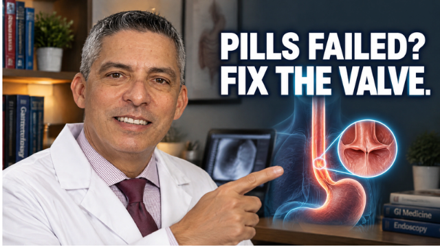
- Antireflux surgery (laparoscopic or robotic fundoplication) is a definitive option for patients with objectively documented, severe or recurrent erosive disease, those who do not wish to take lifelong medication, and those with a significant hiatal hernia or regurgitation-predominant symptoms; it can heal erosive disease by restoring the antireflux barrier (see our antireflux surgery and Nissen Fundoplication pages) (Fass, New England Journal of Medicine, 2022). Referral is appropriate for persistent, severe symptoms despite lifestyle change and at least 6 months of medical therapy, after evaluation with endoscopy, pH monitoring, and high-resolution manometry (Thrumurthy et al., JAMA, 2026).
- Magnetic sphincter augmentation (LINX) and transoral incisionless fundoplication (TIF) are additional options in appropriately selected patients (see our LINX and TIF pages) (Thrumurthy et al., JAMA, 2026).
- Endoscopic dilation is used to treat peptic strictures that complicate erosive disease (Richter and Rubenstein, Gastroenterology, 2018).
Evaluation is warranted for reflux symptoms occurring two or more days a week, for symptoms that persist despite over-the-counter acid suppression, and promptly for any alarm feature — difficulty or painful swallowing, unintentional weight loss, vomiting, or signs of gastrointestinal bleeding such as black stools or anemia (Katz et al., ACG Clinical Guideline, 2022). Because up to half of patients with erosive esophagitis may have few or no typical symptoms, anyone with risk factors (obesity, hiatal hernia, long-standing reflux, older age) and new swallowing difficulty should be evaluated (Katzka et al., Clinical Gastroenterology and Hepatology, 2020).
Referral for a dedicated antireflux evaluation is appropriate for patients with severe (LA grade C or D) or recurrent erosive esophagitis, those whose erosions persist despite optimized PPI therapy, those who prefer not to remain on lifelong medication, and those with a hiatal hernia, stricture, or Barrett's esophagus (Thrumurthy et al., JAMA, 2026). Patients with high-grade disease in particular warrant structured follow-up — including repeat endoscopy to confirm healing and screen for Barrett's — and are often best served by an experienced foregut program that can offer the full range of medical, endoscopic, and surgical therapy (Katz et al., ACG Clinical Guideline, 2022).
Most patients with erosive esophagitis do well long-term, but the disease is chronic and the right long-term strategy depends heavily on grade (Katz et al., ACG Clinical Guideline, 2022). Low-grade disease (LA A and B) can often be managed with the lowest effective PPI dose and may not recur aggressively, whereas high-grade disease (LA C and D) reflects largely irreversible mechanical incompetence of the esophagogastric junction and generally requires either lifelong acid suppression or a definitive antireflux procedure (Katzka et al., Clinical Gastroenterology and Hepatology, 2020). For patients unwilling to commit to indefinite medication, or those with a hiatal hernia or regurgitation-predominant disease, antireflux surgery offers durable control by restoring the reflux barrier (Thrumurthy et al., JAMA, 2026). An integrated foregut program that grades the disease accurately, confirms healing in severe cases, manages weight as the key modifiable factor, and screens appropriately for Barrett's esophagus produces the best long-term results (see our GERD / Acid Reflux pillar).
The prognosis of erosive esophagitis is generally favorable, particularly for low-grade disease. PPI therapy heals erosive esophagitis in roughly 90–95% of patients, and in natural-history studies 73–82% of patients had healed esophagitis at follow-up endoscopy (Mittal and Vaezi, New England Journal of Medicine, 2020; Bi et al., Digestive Diseases and Sciences, 2020). The course tends to be determined early: patients generally continue with the same phenotype over time rather than relentlessly progressing, and a large veteran database study of uncomplicated erosive esophagitis found no patients developing complications over a mean of 4.2 years (Moayyedi and Talley, Lancet, 2006). Progression to Barrett's esophagus occurs in only a minority, regardless of esophagitis severity (Bi et al., Digestive Diseases and Sciences, 2020). The main caveat is severity-dependent recurrence: LA grade C and D disease relapses in nearly 100% of patients within 6 months of stopping therapy, which is why indefinite acid suppression is recommended for high-grade disease (Katz et al., ACG Clinical Guideline, 2022).
Erosive esophagitis is where accurate grading and the right long-term plan make all the difference — low-grade disease can be over-treated, while severe LA grade C and D disease is too often left on inadequate therapy or never evaluated for the hiatal hernia and Barrett's esophagus that frequently accompany it. Dr. Nestor de la Cruz-Muñoz, MD, FACS, DABOM, is a board-certified minimally invasive foregut and bariatric surgeon in Miami with extensive experience across the full spectrum of antireflux therapy — laparoscopic and robotic fundoplication, magnetic sphincter augmentation (LINX), transoral incisionless fundoplication (TIF), hiatal and paraesophageal hernia repair, and Roux-en-Y gastric bypass as an antireflux operation. His practice emphasizes complete evaluation — endoscopy with precise Los Angeles grading, confirmation of healing in severe disease, Barrett's screening, and, when surgery is considered, ambulatory reflux monitoring and high-resolution manometry — so that each patient's treatment is matched to the true severity and mechanism of their disease. Patients benefit from an integrated foregut program, evidence-based medical management, and structured long-term follow-up.
Featured Procedures


FAQ
Most erosive esophagitis heals within about 8 weeks of acid-suppression therapy, and multiple trials confirm that an 8-week course of a proton pump inhibitor achieves mucosal healing in the majority of patients (ASGE Standards of Practice Committee, 2015). Healing is the rule, not the exception — in one large natural-history study, roughly 73–82% of patients had healed esophagitis on follow-up endoscopy, and none progressed to a more severe grade (Bi et al., Digestive Diseases and Sciences, 2020). Recurrence, however, is common once treatment stops: without maintenance therapy, relapse rates reach up to 80% within 3–6 months, which is why severe (LA grade C or D) disease generally warrants indefinite PPI therapy or antireflux surgery (Katz, Journal of the American Geriatrics Society, 1998; Talley and Zand Irani, Journal of Internal Medicine, 2021). Milder grade A/B disease can sometimes be maintained with on-demand or step-down therapy (Okanobu et al., Digestion, 2023).
Erosive esophagitis is a risk factor for Barrett's esophagus, but progression is uncommon and the overall course is benign for most patients. In long-term follow-up, only a minority of patients with erosive esophagitis developed Barrett's esophagus, with similar rates between mild (LA grade A/B) and more severe (grade C) disease (about 5% vs. 14%), and no patients progressed to more severe grades of esophagitis over time (Bi et al., Digestive Diseases and Sciences, 2020). Because severe esophagitis can hide underlying Barrett's, ACG guidelines recommend a follow-up endoscopy after PPI treatment in patients with LA grade C or D disease to confirm healing and evaluate for Barrett's esophagus (Katz et al., American College of Gastroenterology, 2022). It is worth noting that acid-suppression medication heals the mucosa but has not been shown to prevent Barrett's esophagus or reduce cancer risk, which is why durable reflux control and appropriate surveillance matter (Shaheen and Ransohoff, JAMA, 2002).